Moment...


Latest news

ALL ABOUT YOUR HEALTH!

We are determined to keep you regularly informed about our activities. Our Videos are meant to educate you more about your health! Karibu

Dokis-1Gall-Bladder-2.jpg
11/Jul/2024

Cholangitis: is an inflammation of the bile duct system. The bile duct system is found in the liver and is responsible for the transportation of bile from the liver, or to be precise, from the gall bladder, to the first part of the intestine, which is called the duodenum. Bile juice is made in the liver and stored in the gallbladder. In times of digestion, it’s then transported to the duodenum for the digestion of fats and small cholesterol.

Cholangitis is mostly caused by bacterial infections, and in other circumstances, it could be a chronic infection as the result of an autoimmune condition. An autoimmune condition is when the body identifies its own cells as foreign cells, hence attacking them.

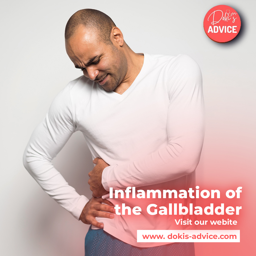
Cholecystitis: this is an inflammation of the gallbladder either due to blockage of the bile duct system with a gallstone or bile sludge. Gallstones are small stones composed of cholesterol that usually form in the gallbladder. Gallstones are a common condition that affects nearly one in every 10 adults.

In medicine, we differentiate:

  1. Calculous cholecystitis: this is when the opening of the gall bladder, that is the cystic duct gets blocked by a gun stone or the bile sludge. This blockage then leads to a buildup of bile juice in the gall bladder, hence increasing the pressure inside, which can lead to inflammation.
  2. Acalculus cholecystitis: this is a guy bladder inflammation that is not caused by gallstones. This can be caused by severe diseases, operations, or traumas.
  3. Chronic cholecystitis: this is a result frequent acute cholecystitis which has been treated using conservative measures like giving antibiotics or healing spontaneously, hence leading to scarring of the gall bladder walls.

There are six major risk factors for the formation of gall stones, which include:

  • Being overweight
  • Women are more affected.
  • Fertile
  • Fair meaning white people or people with a fairly bright skin.
  • Age: around 40 years
  • When it occurs mostly in the family, the risk of other family members getting it is higher.

Cholecystolithiasis: simply refers to having gallstones in the gallbladder.

Choledocholithiasis: this is a term that refers to having gallstones in the bile duct system.

There are some underlying factors that can lead to more frequent building of gallstones. These factors include:

  • Disruption of the enterohepatic circulation: This can be caused by diseases such as Crohn’s disease or after recession of the terminal ileum which is the last part of the small intestine which can lead to bile acid loss syndrome.
  • Fasting, rapid weight loss, parenteral nutrition, or bariatric surgery
  • Haemolytic anaemia: a frequent destruction of red blood cells can lead to increased bilirubin, which can therefore lead to formation of pigment stones from the bilirubin. Bilirubin is a yellow substance that is made after the breakdown of old red blood cells.
  • Hyperparathyroidism: The condition that can lead to excessive calcium levels In the bloodstream, therefore contributing to the formation of gallstones out of calcium.
  • Caroli syndrome: this is a congenital disease that causes the deletion of the bile ducts in the liver intense can contribute to the formation of gallstones.
  • Gilbert syndrome: this is a genetic disorder of bilirubin metabolism that can increase the risk of gallstone formation.
  • cystic fibrosis: The condition that leads to a production of a thick mucus layer that can block the bile ducts hence leading to the formation of gallstones.

Let’s have a look at the signs and symptoms:

  • Nausea and vomiting
  • upper right abdominal pain
  • Painful cramps in the right upper abdominal region
  • Pain projection in the right shoulder
  • Fever
  • Jaundice
  • Itchiness

-Let look at how are these diseases diagnosed:

  • Your doctor will do a physical examination and take your medical history.
  • blood tests: Timeline whether the inflammation parameters are high and parameters of gallbladder obstruction, Liver and Pancreas.
  • Abdominal ultrasound: this is used to determine the structure of the gallbladder, to see if it is filled with gallstones, and to examine the anatomy of the liver.
  • endoscopic ultrasound: this can be used to detect small gallstones in the building system.
  • ERCP/MRCP: the former can be used for diagnostic and therapeutic purposes. The latter can only use for diagnostic purposes. This examination is used to showcase the biliary system. In cases of stenosis, the ERCP can be used for therapeutic purposes, for example, dilation of the biliary tracts, removal of gallstones and stenting. (This is a procedure where one can Insert a plastic or metal hollow tube to help relieve obstruction in areas of stenosis).
  • CT-Scan: mostly for purposes of better operation planning.
  • Abdominal X-Ray: done in suspicion of a hole or tear in the gall bladder.

Now let’s delve into how the above diseases are treated:

  • giving pain medications,
  • giving medication against cramping
  • Not eating or drinking to relieve the strain of the gallbladder.
  • Giving the patience fluids directly into the vein to prevent dehydration.
  • in cases of high inflammation parameters, giving antibiotics
  • Surgery for the removal of the gallbladder may be recommended after initial treatments to prevent acute cholecystitis and for the prevention of potential complications.
  • In other instances, surgery may not be recommendable, hence the importance of an ERCP, as stated in the diagnostics above.

So, you may ask yourself, are there ways to reduce the risk of getting cholecystitis?

  • Eating a healthy balance today while reducing the number of high cholesterol foods
  • Regular exercise reduces the risk of being overweight or obese.
  • avoiding Rapid weight loss diets

remember to always seek medical advice.


Alcoholism-Liver-cirrhosis-1.png
01/Sep/2023

According to WHO data in 2020 Deaths caused by liver diseases in Kenya were about 13,400 or 5.09% of the total deaths. With an age rate of 57.44 deaths per 100,000 of the population, ranking Kenya at number 6 in the world.

Leading causes of death in Kenya as per worldlifeexpectancy.com sept/2020

  1. Stroke: 11,453 deaths
  2. Influenza and pneumonia:16,263
  3. Coronary Heart diseases:8,626
  4. Liver diseases:9,658
  5. TB:14,162
  6. HIV/AIDS:15,048
  7. Road accidents:10,723
  8. Malaria: 9,116
  9. Diabetes mellitus:4,072
  10. Hypertension:3,183
  11. Lung diseases:3.084
  12. Cervical cancer:2,441
  13. Prostate cancer: on this date no Data
  14. Kidney diseases:3,216

Liver cirrhosis is a condition where the liver becomes scarred and damaged over time, which can prevent it from performing its important functions. Let’s break down these functions in a simpler way:

  • Detoxification: The liver helps remove harmful substances like drugs, alcohol, and ammonia from the body.
  • Metabolism: It converts certain hormones and substances into forms that the body can use. For example, it activates a thyroid hormone called T4 into its active form, T3.
  • Cholesterol production: The liver produces cholesterol, which is important for various functions in the body.
  • Blood sugar balance: It helps regulate the amount of sugar (glucose) in the bloodstream by removing excess glucose and storing it as glycogen.
  • Bile production: The liver produces bile, a substance that aids in the digestion of fats.
  • Protein synthesis: It creates important proteins like albumin, which is crucial for maintaining proper blood circulation, and factors needed for blood clotting.
  • Blood filtration: The liver filters the blood, removing toxins, by-products, and other harmful substances.

In liver cirrhosis, the liver’s ability to perform these functions is compromised due to the scarring and damage it has undergone.

Liver cirrhosis can have various causes. Here are some common ones:

  • Excessive alcohol consumption over many years can lead to liver cirrhosis.
  • Virus infections can also cause liver cirrhosis, such as:
  • Hepatitis A: Contracted through contaminated food or drinks, especially seafood, or direct contact with an infected person. However, it usually doesn’t lead to cirrhosis as most people recover fully and develop lifelong immunity.
  • Hepatitis B and C: Transmitted through sexual intercourse with an infected person, sharing needles, or from mother to child during pregnancy or delivery.
  • Hepatitis E: Ingestion of contaminated water containing fecal matter from infected individuals. This type of hepatitis is typically self-limiting and doesn’t usually result in cirrhosis.
  • Hepatitis D: Often occurs as a co-infection with Hepatitis B.
  • Non-alcoholic steatohepatitis (NASH): This condition is characterized by the buildup of excess fat in the liver and is commonly seen in individuals with diabetes mellitus.

Liver cirrhosis can be classified as either:

  • Compensated: This is when one suffers from cirrhosis but has no acute symptoms like the one’s that will be discussed below.
  • Decompensated: This is when the listed below symptoms occur.

The signs and symptoms of liver cirrhosis can vary, but here are some common ones:

  1. Loss of appetite
  2. Weight loss and muscle wasting
  3. Nausea and vomiting
  4. Itchy skin and jaundice (yellowing of the skin and eyes) due to elevated levels of bilirubin in the blood.
  5. Appearance of tiny red lines on the skin
  6. Hair loss, particularly noticeable in men who lose their chest hair.
  7. Swelling of the legs, ankles, and feet (oedema) caused by fluid retention.
  8. Abdominal swelling due to the accumulation of fluid known as ascites.
  9. Frequent or prolonged bleeding due to a deficiency of clotting factors produced by the liver.
  10. In advanced stages of cirrhosis, complications may arise, including:
  • Vomiting of blood due to bleeding from enlarged blood vessels in the gastrointestinal tract.
  • Increased pressure in the portal vein, leading to the diversion of blood flow into smaller blood vessels that may rupture under high pressure.
  • Kidney failure, liver failure, the development of hepatocellular carcinoma (liver cancer), and ultimately, death.

It’s important to note that liver cirrhosis is a serious condition that requires medical attention and treatment.

The treatment and interventions for liver cirrhosis include:

  • Lifestyle changes: It is crucial to make certain lifestyle modifications to manage liver cirrhosis effectively. These include avoiding alcohol consumption, quitting smoking, maintaining a healthy weight (especially in cases of obesity or overweight), engaging in regular exercise, and getting vaccinated against hepatitis A and B.
  • Medication: Medical treatment for liver cirrhosis should always be discussed and prescribed by a healthcare professional. Some medications commonly used in cirrhosis management include:
  • Diuretics: These medications help relieve swelling (oedema) and ascites by promoting the removal of excess fluid from the body.
  • Beta-blockers: Drugs like propranolol can be prescribed to reduce portal hypertension, which is high blood pressure in the portal vein that supplies the liver.
  • Antibiotics: In case of bacterial infections, antibiotics may be administered to prevent or treat complications.
  • Diabetes treatment: If diabetes coexists with cirrhosis, appropriate management of blood sugar levels is important.
  • Laxatives: Laxatives may be prescribed to help with the treatment of hepatic encephalopathy, a condition caused by the accumulation of ammonia in the bloodstream due to impaired detoxification by the liver.
  • Endoscopic treatment: In cases of variceal bleeding in the gastrointestinal tract or stomach, endoscopic procedures can be performed to stop the bleeding. This may involve the use of techniques like banding or sclerotherapy to close off the bleeding vessels.
  • Liver transplant: In severe cases where the liver is extensively damaged and other treatments are ineffective, a liver transplant may be considered. This involves replacing the diseased liver with a healthy liver from a donor.

Diet for cirrhosis

Therapeutic role of nutrition in liver cirrhosis has been known for many years and as such, nutritional status was one of the criteria observed in the initial prognostic tests for liver disease.

Being malnourished is also very common in people with cirrhosis with about 20% of people with compensated cirrhosis being malnourished and more than 50% of people with decompensated cirrhosis. When dealing with liver cirrhosis, being malnourished is a risk factor for succumbing to the disease. So, to enable better coping, reversal of malnutrition to a state of good nutrition is key.

In general, a diet that ameliorates or prevents the symptoms of liver cirrhosis as described above is a good diet for management. This is for example a diet that prevents high pressure in veins, a diet rich in albumin etc.

Some of the diet recommendations are:

  • Eat little and often to prevent the body from being stressed and breaking down the proteins in your muscles.
  • Eat more calories and protein because when cirrhosis happens, your liver is not able to store glycogen which the body needs so you compensate by eating more.
  • Reduce salt to help manage fluid retention and bloating (ascites and oedema) as salt can worsen fluid intake and swelling in the liver.
  • Managing hyperglycaemia (high blood sugar) and diabetes as liver plays a major role in managing blood sugar but then with cirrhosis, the capacity is impaired.
  • Supplements for people with or at risk of bone disease (osteoporosis) because factors that contribute to bone loss need to be reduced when with cirrhosis
  • Coffee intake as coffee helps in breaking down fats.
  • High intake of fruits and vegetables

It’s important to consult with a healthcare professional for an accurate diagnosis, appropriate treatment options, and ongoing management of liver cirrhosis.

And as always keep yourself informed at Doki’s Advice


04/Feb/2025

Understanding Chronic and Coronary Heart Disease:

What are Chronic Heart Disease and Coronary Heart Disease?

Chronic heart disease (CHD), often referred to as cardiovascular disease, is a long-term condition that affects the heart and blood vessels. It commonly results from narrowed or blocked arteries, leading to reduced blood flow to the heart. This can increase the risk of heart attacks, heart failure, arrhythmias, and other complications. CHD is a leading cause of death worldwide and requires long-term management to prevent severe outcomes.

Coronary heart disease, also known as ischemic heart disease, is a specific type of chronic heart disease that results from plaque buildup in the coronary arteries. This buildup restricts blood flow to the heart muscle, leading to conditions such as angina (chest pain) and heart attacks. While all coronary heart disease is chronic, not all chronic heart disease is limited to coronary artery issues; other types may include heart valve disease and arrhythmias.

Types of coronary heart diseases:

  • Acute coronary disease (ACS): sudden heart problems, like heart attacks
  • Chronic coronary Syndrome (CCS): Long-term or stable heart related issues
  • Asymptomatic CHD: No symptoms despite presence of the disease

How are Chronic and Coronary Heart Disease Identified and Diagnosed?

The diagnosis of these conditions involves several steps, including medical history assessment, physical examination, and diagnostic tests. Some of the common ways to identify CHD and coronary heart disease include:

Symptoms – These conditions often present with symptoms such as:

  1. Chest pain or discomfort (angina)
  2. Shortness of breath either at rest or during physical activity
  3. Fatigue
  4. Swelling in the legs, ankles, or feet
  5. Irregular heartbeat


Medical Tests – To confirm a diagnosis, doctors may use:

  • Electrocardiogram (ECG/EKG): Measures electrical activity in the heart to detect irregularities.
  • Echocardiogram: Uses ultrasound to visualize heart function and structure.
  • Stress Test: Evaluates how the heart performs under physical exertion.
  • Coronary Angiography: Uses contrast dye and X-rays to detect blockages in the coronary arteries.
  • Blood Tests: Check cholesterol, blood sugar, and markers of heart disease.
  • Cardiac catheterisation

How are Chronic and Coronary Heart Disease Treated?

The treatment of these conditions depends on the severity of the disease and the patient’s overall health. Treatment options include:

Lifestyle Changes:

  • Eating a heart-healthy diet rich in fruits, vegetables, whole grains, and lean proteins.
  • Engaging in regular physical activity (at least 30 minutes a day, most days of the week).
  • Maintaining a healthy weight.
  • Avoiding smoking and excessive alcohol consumption.
  • Managing stress through relaxation techniques and adequate sleep.


Medications:

  • Statins: Lower cholesterol levels to prevent artery blockages.
  • Beta-blockers: Reduce heart rate and blood pressure to ease strain on the heart.
  • ACE Inhibitors & ARBs: Help relax blood vessels to improve blood flow.
  • Aspirin or Antiplatelet Drugs: Prevent blood clot formation.
  • Diuretics: Reduce fluid buildup in patients with heart failure.
  • Long acting-nitrates: Dilating coronary arteries and reducing preload hence improving oxygen supply to the heart.
  • Ranolazine: reduce myocardial oxygen demand (this is just a symptomatic therapy).
  • Ivabradine: reduces heart rate without affecting blood pressure or contractility.
  • Blood thinners

Medical Procedures and Surgery:

  • Angioplasty and Stenting: A minimally invasive procedure to open blocked arteries.
  • Coronary Artery Bypass Grafting (CABG): A surgical procedure that improves blood flow to the heart by bypassing blocked arteries.
  • Implantable Devices: Pacemakers or defibrillators may be used to manage arrhythmias.


How to Prevent Chronic and Coronary Heart Disease

  • Preventing heart disease is possible by making healthy lifestyle choices and managing risk factors. Key preventive measures include:
  • Maintain a Healthy Diet: Focus on foods rich in fiber, healthy fats, and low in processed sugars and sodium.
  • Exercise Regularly: Engage in activities like walking, swimming, or cycling to improve cardiovascular health.
  • Control Blood Pressure and Cholesterol: Regular check-ups and necessary medications can help manage these risk factors.
  • Quit Smoking: Smoking damages blood vessels and significantly increases heart disease risk.
  • Manage Diabetes: Keeping blood sugar levels under control reduces damage to the heart and blood vessels.
  • Reduce Stress: Practice mindfulness, yoga, or other relaxation techniques to lower stress levels.


Regular Medical Check-ups: Routine screenings help detect early signs of heart disease and take preventive action. And as usual always seek medical advice from your doctor first.


MeningitisMeningitis-2.jpg
07/Jun/2024

So, what do we understand by this?

Meningitis is an inflammation of the protective layer membrane that covers the brain and the spinal cord. Anyone can get meningitis, from small babies to older people, and it can be very severe, causing brain and spinal cord damage and long lasting disabilities.

Meningitis is caused by various agents, like bacteria (Neisseria meningitidis, pneumococcal bacteria, and bacteria causing tuberculosis), viruses such as HSV, chickenpox, and other harmless viruses. An infection caused by the HSV, chicken pox viruses, or bacterial meningitis is considered an absolute emergency that needs immediate treatment with antiviral or antibacterial drugs. Meningitis caused by bacteria can be treated by giving antibiotics, depending on the  bacteria that caused the meningitis in the first place, and it should be an antibiotic that can pass through the brain-brain barrier.

Before we delve into the topic, it is important to state that, meningitis can be prevented through vaccination. The meningitis vaccine can be given to children from the age of 12 months and all other age groups, especially those living within the meningitis belt. So, what exactly is the meningitis belt? These are the regions in Africa that are highly affected by meningitis, hence, a recommendation for the meningitis vaccine for kids of the age of 12 months as well as a pneumococcal vaccine.

Meningitis is spread by respiratory secretions through sneezing, coughing or kissing. In some cases, we do have meningitis carriers who can spread the infection yet they don’t suffer from it.

Children are mostly affected by bacterial variants, for example, the pneumococcal variant, which is also the leading cause of pneumonia.

What are some of the signs and symptoms of meningitis?

  • Headaches
  • high fever
  • nausea and vomiting
  • irritability to light
  • confusion
  • seizures
  • paralysis
  • Drowsiness or unresponsiveness
  • stiff neck
  • a rash that does not fade

The symptoms do not appear in any order and to some extent, some patients do not show any of the of the signs or symptoms stated above.

The incubation period for bacterial meningitis is around 2  to approximately 4 days but can also extend to 10 days. It is very important to recognise bacterial meningitis at an early stage and begin with antibiotic medication, as it is crucial because it can quickly end in death within hours or days.

The incubation period for viral meningitis is around 2 to 14 days. It mostly begins acutely, lasting a few hours to a few days. HSV or chickenpox virus caused meningitis, also needs early therapy with antiviral drugs. Other less harmless viruses that cause meningitis can heal by themselves after a couple of days to weeks. But since we cannot differentiate which viral infection or bacterial infection is causing meningitis, it’s always advisable to seek medical advice.

Let’s delve into the diagnosis of meningitis

  • clinical examination through your physician
  • inspection of your skin
  • examination of the meningitis sign
  • blood tests and microscopic examination of a blood stain
  • Lumbar puncture to collect cerebrospinal fluid for microscopic examination
  • in some cases, a CT-Scan can be necessary before the lumbar puncture. This is important in situations where symptoms such as unresponsiveness, loss of consciousness neurological symptoms that show suspicion of intracranial pressure signs are present.

So how do you treat a meningitis?

  • Early antibacterial treatments
  • supplementing the treatment with the antiviral treatments by suspicion of viral meningitis
  • cortisone in cases of intracranial pressure suspicion
  • supportive therapy such as
  • giving paracetamol or ibuprofen for pain or fever’s
  • Therapies for seizures in cases where epileptic seizures have been reported.
  • giving blood thinners in cases of septic thrombosis

Exposed people to those affected with bacterial meningitis are also given a prophylactic antibacterial medication to help reduce the Spread.

It is crucial to always begin an early antibacterial or antiviral therapy because if meningitis is not treated properly or early enough it could lead to severe long-term problems such as:

  • Hearing or vision losses
  • problems with memory and concentration
  • recurrent seizures afterwards
  • problems with coordination, movement, and balance
  • loss of limbs due to necessity of an amputation of affected limbs

Always seek medical advice and remember prevention is better than cure, so do get vaccinated as well as your children.


Cervical-cancer-doc.jpg
13/May/2024

Cancer of the cervix happens when normal cells in the cervix change into abnormal cells and grow out of control. Most people whose cervical cancer is found and treated early do very well. The cervix is the bottom part of the uterus, where it meets the vagina. The cervix is composed of two main types of cells. The outer layer of the cervix is covered with cells called squamous cells. Squamous cell carcinoma of the cervix is the name for a cancer that affects these cells. The cervix also includes glandular cells, which line the canal of the cervix that leads into the uterus. These cells can also become cancerous; when they do, they are called adenocarcinoma of the cervix. Although they arise from different types of cells, squamous cell carcinoma and adenocarcinoma of the cervix are treated similarly in the early stages.

Risk Factors

Most cervical cancers are caused by infection with a virus called human papillomavirus. HPV is spread by direct skin-to-skin contact, includi ng sexual intercourse, oral sex, anal sex, or any other contact involving the genital area (eg, hand-to-genital or mouth-to-genital contact). HPV infection can also cause a noncancerous condition called condyloma, which is caused by genital warts.

HPV infection is very common. Approximately 75 to 80 percent of sexually active adults will acquire a genital HPV infection before the age of 50. There are many types of HPV, and these affect different areas of the body. Most HPV infections are temporary because the body’s immune system effectively clears the infection.

Risk factors for cervical cancer include:

  • Smoking tobacco
  • Increasing number /constant change of sexual partners. The greater your number of sexual partners and the greater your partner’s number of sexual partners, the greater your chance of getting HPV.
  • Early sexual activity. Having sex at an early age increases your risk of HPV.
  • Bad sexual hygiene
  • Other sexually transmitted infections.
  • A weakened immune system, e.g HIV/AIDS
  • Genetic prädisposition
  • Exposure to miscarriage prevention medicine. If your parent took a medicine called diethylstilbestrol, also known as DES, while pregnant, your risk of cervical cancer might be increased. This medicine was used in the 1950s to prevent miscarriage. It’s linked to a type of cervical cancer called clear-cell adenocarcinoma.

Symptoms

Typically, cervical cancer develops slowly over several years. In some cases, the cancer does not cause any symptoms, while in others, it causes abnormal vaginal bleeding or discharge. This can include vaginal bleeding between menstrual periods, bleeding after sex, or bleeding after menopause. This bleeding may be spotting or heavy bleeding.

Other symptoms may include:

  • Menstrual bleeding that is heavier and lasts longer than usual.
  • Watery, bloody vaginal discharge that may be heavy and have a foul odor.
  • Pelvic pain or pain during intercourse.

Diagnosis

Pap testing and human papillomavirus (HPV) testing are commonly used together to screen for cervical cancer. If a Pap test shows abnormal cells, further testing is essential, as treatment of abnormal cells of the cervix can prevent cervical cancer. A biopsy of the cervix involves removing a small piece of tissue from the cervix. The biopsy is performed during an office visit using a procedure called colposcopy. The colposcope, which is similar to a large magnifying lens, magnifies the view of the cervix. This allows the clinician to better see the location, extent, and degree of cervical abnormalities that may not be visible with the naked eye alone.

The tissue obtained during the biopsy is examined with a microscope to see if cervical cancer cells are present. In some cases, more of the cervix will need to be removed for biopsy; this is done through a procedure called cervical conization or a cone biopsy. This can be done either in the operating room by a surgeon using a scalpel or in the office using a loop electrosurgical excision procedure (LEEP). LEEP is performed with a device that uses an electric current to remove a piece of the cervix. You will usually get the results of the biopsy one to two weeks after the biopsy is done.

CT-scan or MRI of the Pelvis Region: to determine spreading of the tumor to other organs or regions of the body.

Treatment

Cervical cancer can be treated in different ways:

  • Surgery: Some cases of cervical cancer are treated with surgery to remove the cancer. Different types of surgery can involve:
  • Removing the cervix, uterus, and upper part of the vagina is called a “radical hysterectomy.”
  • Removing all or part of the cervix but leaving the uterus in place—this surgery is done only in special situations.
  • Radiation therapy: Radiation kills cancer cells.
  • Chemotherapy: Chemotherapy is the medical term for medicines that kill cancer cells or stop them from growing. People with cervical cancer usually receive chemotherapy at the same time as radiation therapy.

Prevention

Cervical cancer can be prevented through:

  • HPV Vaccination.
  • Routine Pap tests.
  • Practicing safe sex.
  • Proper Hygiene
  • Avoid cigarette smoking.

Dokis-Pneumonia-2-1200x1200.jpg
04/Apr/2024

Pneumonia

Pneumonia is an inflammation of the lungs that is mostly caused by bacterial infections but can also be caused by viral infections. It is the most common reason for deaths in developing and in industrial nations.

In Kenya, pneumonia is responsible for approximately one in five children’s deaths. It claims approximately 8,000 to 10,000 children’s lives annually.

Pneumonia takes about two to four weeks before healing. For small children and older people people with lung and heart conditions, the infection will take longer and even probably require hospitalization due to the severity of their conditions.

As mentioned earlier, pneumonia is caused by vectors such as bacteria and viruses, and in patients with a reduced immune system, pneumonia can also be caused by fungi.

Some of the factors that increase susceptibility to pneumonia infections include:

  • age factors such as young babies and older people
  • seasonal factors like rainy and cold seasons
  • Prior medical history such as asthma in COPD (chronic obstructive pulmonary disease),

Pneumonia can be categorized into:

  • community-acquired pneumonia: the most common bacteria are pneumococcal, and Haemophilus influenza, others include: Chlamydophila pneumococcal, Mycoplasma pneumococcal, Legionella, Staphylococcus aureus, and Mycobacterium tuberculosis.

-Viruses such as: Influenza, RSV, and Corona Virus

  • Hospital-acquired pneumonia: the most common bacteria for these are Pseudomonas aeruginosa and Staphylococcal.
  • Pneumonia on patients with a reduced immune system.

mostly caused by minor bacteria and fungi

  • Neonatal Pneumonia

Let’s look at signs and symptoms of pneumonia:

  • Fever
  • Coughing up yellow or green mucus
  • Chest pains and pains while breathing
  • Aching of the body, joints and headaches
  • Exhaustion
  • Loss of appetite
  • Wheezing while breathing
  • Confusion and hallucinations in older patients or due to certain viral infections like influenza

Now that we know the signs and symptoms to look out for, let’s dive into how we diagnose pneumonia:

  • Clerking: this is when the doctor asks general questions about your cough, including when the symptoms started and any prior medical history
  • the doctor checking for signs like strenuous breathing and blueness of the lips and tongue, this is due to less oxygen intake in the lungs.
  • The doctor will do a physical examination using his stethoscope to determine the different sounds of the lungs while breathing
  • Blood tests: this helps to determine between a viral and a bacterial infection. In the case of a bacterial infection, the white blood cells and inflammation parameters are mostly high. In the case of a viral infection, the white blood cells and inflammation parameters will be normal or just very slightly elevated. In other cases, one could have both a viral and a bacterial infection, this is what you would call a superinfection in medical terms.
  • X-ray: this helps to determine where exactly the pneumonia is located; for example, it could be on both sides of the lungs or just on one side.

Now that we know the different diagnostic steps, let’s have a look at how he treats pneumonia.

  • Bacterial infections are always treated with antibiotics.
  • Viral infections do not require antibiotics. Mostly, this kind of pneumonia just requires anti-inflammatory treatment with medications such as Ibuprofen, and paracetamol, and enough fluid intake.
  • In case of a superinfection with a bacterium and a virus, antibacterial therapy will be required.
  • In case of a fungal infection, antifungal therapy is required.

Please note that not all kinds of pneumonia require antibiotics, hence the need to draw blood to differentiate between a bacterial, viral, or fungal infection. This is crucial to avoid the high rise of resistance in antibiotic therapies.

We do say prevention is better than cure, so how can we reduce the frequency in which pneumonia occurs in our general population? The main method of pneumonia prevention, especially in small children and older patients is vaccination.

-small babies and children can be vaccinated against Haemophilus and pneumococcal disease at the age of two months. Older patients as well as patients with a reduced immune system can also prevent the severity of pneumonia by getting vaccines against the named bacteria above.

For more medical gems, keep at Doki’s Advice.


Dokis-colorectal-2.jpg
17/Mar/2024


This is a malignant tumour found in the lining of the large intestines up to the rectum.

The large intestine is known for its role in the digestive system to absorb water and remove waste from the food we eat.

Colon and rectal cancer mostly occur at older age, but by a particular number of people who have certain gene mutations, this cancer can occur at an early age, for example, at 30 without them realising it.

Colorectal cancer is the third most common cancer in the world and the second leading cause of death globally.

According to the data required from Globocan, there were an estimate of 1,354 new cases and 937 deaths from colorectal cancer in Kenya in 2018. Other data sources have noticed that there is a rise in colorectal cancer cases in Kenyan rural areas and that it is the fifth most common cancer in sub-Saharan Africa.

Let’s delve into factors that increase the risk of colorectal cancer:

  • Genetic Factors:
    • Such as familial disposition
    • Familial polyposis syndrome: Families with this syndrome tend to have the growth of many polyps along the large intestines at a very young age. These polyps can with time, go through genetic mutations, causing them to be cancerous and hence leading to early-stage colorectal cancer. To note, with this syndrome, the growth of polyps can also occur in any part of the intestines.
    • Hereditary nonpolyposis colorectal cancer syndrome: This is also another genetic disease that increases the risk of colorectal cancer.
  • Eating habits and lifestyle:
    • Smoking and excessive drinking of alcohol.
    • Obesity
    • Low fibre or high fat diet
    • Red meat or processed meat and foods

-Some protective factors would be:

  • Exercise
  • Eating foods rich in fibre, vegetables, and less meat
  • Maintaining healthy weight

Now that we know the risks and protective factors, let’s have a look at the signs and symptoms of colorectal cancer:

  • Most of the time, there are no characteristic early symptoms
  • A constant change of bowel movements from Obstipation and diarrhoea
  • Rectal bleeding: this can either be visible blood or non-visible, meaning it can only be detected by doing a faecal occult blood test.
  • Persistent abdominal discomforts such as cramps passing gas or pain
  • Unexplained weight loss within a short period of time
  • Feeling like the bowl isn’t completely empty even after passing stool.
  • Fatigue and weakness
  • Unexplained Anaemia


So how do we diagnose colorectal cancer?

  • A basic diagnostic is performing a digital rectal examination from your doctor. This is used to detect palpable Tumours just near or around the rectal and anal regions.
  • Complete colonoscopy with biopsy: This is an examination of the whole large intestine with the possibility of taking biopsy samples to pathologically test the sample for tumours type.
  • Rectoscopy for rectal cancer diagnosis.
  • CT-Scan for staging purposes: is the tumour metastasized? if yes to what organs.
  • Lab tests for tumour markers such as CEA as a control value especially while doing chemotherapy and years after remission.
  • MRI-Scan: mostly in regard of rectal cancer.

After a proper diagnosis of colorectal cancer the next step would be to discuss with your oncologist and surgeon what the individual therapy plan will look like. This is determined by factors like

  • how physically fit is the patient prior to therapy,
  • is the patient eligible for surgery or not,
  • size of the tumour
  • is the tumour only at one spot or several spots in the bowel?
  • does the tumour have certain genetic changes?
  • Is prior Radio-chemotherapy necessary or not.

-Let have a look at the different therapy options

  • Surgery: This is the option for tumours that are just locally on one spot or multiple tumours localised on the same segment of the large intestine. Some tumours could have spread to the liver but could still be operable when the metastasis is either singular or multiple but localised in the same area. Such factors would have to be considered by your surgeon.
  • Chemotherapy: Some Tumours could be so large that prior chemotherapy is required to shrink them to an operable size.
  • Radiotherapy: This is mostly done on rectal cancers since colon cancers are insensitive to radiation.

Which therapy regime each patient needs depends on some of the factors noted above. Some may need a Stoma bag after an operation. This will also be discussed with you during the planning stage for the therapy needed.

As always, seek medical advice and keep it Doki’s Advice for more info gems.


Dokis-vertigo-1200x1200.jpg
04/Mar/2024

It does not in totality equate to dizziness but is a form of dizziness accompanied by a whirling, spinning sensation with a lack of postural stability or balance. Can be accompanied with Nausea and Vomiting

Vertigo can be a disease on its own, but it can also be a symptom of a chronic or acute condition.

For a better understanding, let’s remotely understand how balancing works. Balancing involves a complicated connection between the brain, inner ears, eyes, spine, and muscles. Disturbances, injuries, and infections of the above-mentioned can result in dizziness or vertigo.

Causes range from benevolent to life-threatening conditions.


Let’s start with the common ones:

Side effects of Medications such as antibiotics, chemotherapy, etc.

* Specific forms of migraines, like vestibular migraines.

*Motion Sickness.

peripheral Vertigo involving the inner ear (vestibular organ)


Today we focus on peripheral vertigo as a result of malfunctioning of the inner ear.

It’s important to note that the inner ear, being one of the smallest organs, has a complex ib structure with very many microscopic structures coordinating earing and balancing. We will spare you the details of how it works.

Peripheral vertigos include:

  1. Benign paroxysmal positional Vertigo.

The most common vestibular disorders.

Vertigo or dizziness with acute onsets may last a few seconds to a few minutes.

The vertigo manifests itself with a change in body posture: turning in bed, movement of the head.

Trigger in layman’s tongue: irritation of the inner ear due to loose microscopic fragments detaching themselves from their fixation and circulating within the microscopic inner channels of the ear.

The diagnosis is clinical.

Therapy is the repositioning of detached fragments with physio—therapeutic special vertigo training.


2.Neuropathia Vestibularis.

Patients present with severe vertigo, mostly associated with nausea and vomiting and conspicuous loss of balance.

It is believed to be associated with an inflammation of the inner ear.

The diagnosis is also clinical after a thorough examination to rule out possible central causes.

Patients profit from steroids such as prednisolone and other symptomatic medications like vomex.

3.Meniere Disease.

Presents with a triad of tinnitus, loss of hearing, and severe vertigo.


Red flags of acute dizziness or vertigo:

In acute onsets, it is important for a clinician to note that most Vertigo’s are harmless. Underlying disorders may be overseen.

These include strokes of the brainstem, tumors, or even cardiac conditions.

It is important to rule out these conditions.

Using MRI scans. Long-term ECGs.

The most important thing is to always do a thorough clinical examination and a well-sourced patient’s history. When in doubt as a clinician, better do more tests than ignore your instincts.


Lastly, as a patient, before presenting yourself with dizziness, ask yourself simple questions like:

Did I hydrate well? Is my blood sugar okay?

And, as always, seek medical advice for better treatment.


Dokis-Prostate-cancer-1200x1200.jpg
04/Mar/2024

Prostate cancer is one of the most common types of cancer. Prostate cancer occurs in the prostate, which is a wall-nut-shaped gland in males. The function of the prostate is to produce seminal fluid that transports and nourishes sperm. Many prostate cancers grow slowly and are confined to the prostate gland, where they may not cause serious harm. However, while some types of prostate cancer grow slowly and may need minimal or even no treatment, other types are aggressive and can spread quickly. Prostate cancer, when detected early, has a better chance of being successful.

What are the risk factors for prostate cancer?

It’s not clear what causes prostate cancer. However, the following risk factors may play a role:

  • Race: For reasons not yet determined, black people have a greater risk of prostate cancer than do people of other races. In black people, prostate cancer is also more likely to be aggressive or advanced.
  • Obesity: People who are obese may have a higher risk of prostate cancer compared with people considered to have a healthy weight, though studies have had mixed results. In obese people, the cancer is more likely to be aggressive and more likely to return after initial treatment.
  • Older age: Your risk of prostate cancer increases as you age. It’s most common after age 50.
  • Family history: If a blood relative, such as a parent, sibling, or child, has been diagnosed with prostate cancer, your risk may be increased. Also, if you have a family history of genes that increase the risk of breast cancer (BRCA1 or BRCA2) or a very strong family history of breast cancer, your risk of prostate cancer may be higher.


What are the symptoms of prostate cancer?

Prostate cancer may show no signs in its early stages.  When advanced, it may cause the following symptoms:

  • Trouble urinating
  • Decreased force in the stream of urine
  • Blood in the urine
  • Blood in the semen
  • Bone pain
  • Losing weight without trying
  • Erectile dysfunction

What are the complications of prostate cancer?

  • Metastasis: Spread to nearby organs, including the bones and bladder.
  • Urinary Incontinence: Inability to control passage of urine
  • Erectile dysfunction


How is prostate cancer diagnosed?

  • PSA Testing: This is the most commonly used and most valuable test for early detection of prostate cancer. The likelihood of prostate cancer increases with more elevated PSA values.
  • Age-specific ranges include
  • 40 – 49 years (0 to 2.5ng/mL)
  • 50–59 years (0 – 3.5 ng/mL)
  • 60 – 69 years (0-4.5 ng/mL)
  • 70–79 years (0 – 6.5bng/mL)
  • Digital Rectal Examination (DRE): This involves the insertion of the digit finger into the rectum to assess for prostate enlargement. DRE may detect psotate nodules, induration, or asymemetry that can occur with prostate cancer.
  • Transurethral ultrasound is often used to evaluate abnormalities detected on DRE.
  • Prostate Biopsy: Depending on the findings of DRE, PSA, and prostate ultrasound, a biopsy of the prostate may be recommended. Biopsy is done to obtain tissues for histologic diagnosis. This is the definitive diagnosis for prostate cancer.


How is prostate cancer treated?

  • Active Surveillance: This involves routine tests to check whether the cancer is starting to grow more quickly. If so, active treatment should be initiated.
  • Surgery: This involves the removal of the prostate gland through surgery.
  • Radiation Therapy: Radiation kills cancer cells. Radiation can be given from a machine that moves around the body or from a direct source to the prostate gland.
  • Hormone Therapy: The male hormones are responsible for the growth of the prostate gland. Hormone therapy reduces the levels of the hormones and thus shrinks the cancer. Hormone therapy can be done through the removal of testicles or through medications.
  • Chemotherapy: Advanced prostate cancer requires chemotherapy for complete treatment.

Remember to always seek medical advice from your practitioner.


30/Jan/2024

In Kenya, cancer is the 3rd leading cause of death, accounting for approximately 7% of the deaths per year, after infectious diseases and cardiovascular diseases.

Generally, it’s difficult to get accurate data due to circumstances such as:

Low collection accuracy and underdiagnosis. A huge number of cancer patients could go undiagnosed, especially if they lack adequate funds for diagnostics and treatment, hence avoiding going to hospital.

Most of the data collected comes from Nairobi and other urbanised settings.

Most leading cancers in Kenya are:

  • Breast cancer: 34 cases per 100,000 people
  • Cervical cancer: 25 per 100,000 people
  • Prostate cancer: 17 cases per 100,000 people
  • Oesophageal cancer (food gut): 9 per 100,000 people

Approximately 75% of cancer cases are diagnosed in late stages due to factors named above as well as lack of awareness.

So, let’s dive into lung cancer as well as other types of cancers we will handle in coming articles.

Lung cancer is a malignant tumour growth that can occur in either side of the lungs or both. This can either be the main tumour or a metastasis (cancer spreading) of other types of cancers of the prostate, breast, head, neck, kidney, and womb, just to mention a few.

The downside of most cancers is that they have no initial symptoms that one could watch out for, in order to push one to go for an early check-up.

Nonetheless, lung cancer patients could experience common symptoms such as:

  • Persistent dry coughing
  • Coughing up blood
  • Breathlessness or difficulties breathing.
  • Unexplained loss of weight: particularly a huge amount of weight in within a short period of time
  • Signs of anaemia: Loss of breath, getting tired quickly, pale face, heart palpitations
  • Unexpected loss of appetite
  • Pain while coughing or breathing.
  • Problems swallowing
  • Hoarseness of the voice
  • Intermittent night fevers, sweating at night.

There are two main types of lung cancer:

  • Non-small-cell lung cancer: this is the most common form, accounting for about 85% of most cases. It grows slowly before getting to the size in which it could start causing symptoms.
  • Small-cell lung cancer accounts for about 15% or less of lungs cancers. It has a very fast growth rate hence could lead to quick deterioration of a patient’s symptoms due to early metastases (spreading).

The type of lung cancer a patient has determines the kind of therapy they will require.

The age at which most people are affected is around the age of 40 and above. Rarely does it occur to younger patients.

The most common cause of lung cancer, as we all know, is of course, smoking, which accounts for approximately 70% of the cases. This does not mean that nonsmokers cannot develop lung cancer.

Other causes of lung cancer among nonsmokers are:

  • Long exposition to asbestos and radon exposure (a few studies have found high exposure to radon in the coastal region, especially in Taita/Taveta region and around Nairobi Kenyatta University).
  • Exposure to Industrial gases and vehicle exhaust gases
  • Exposure to polycyclic aromatic hydrocarbons: like cooking indoors with wood, there is a constant inhalation of the gases produced. This has also been seen to cause food-gut cancers.
  • Exposure to some metals like: Arsen, chromium or Nickel

Let’s see how lung cancer can be diagnosed:

  • X-Ray: though with this method, it’s rare to be able to see the tumours
  • CT-Scan: This is the ideal method of diagnosis to determine the areas of metastasis, like the bone and brain.
  • Getting a sample of the growth and sending it in for pathological testing to prove if its malignant or not.
  • One can also do lab tests for markers in the blood.

– NSE: Mostly high in small-cell lung cancers

–  LDH: generally, suspiciously high in most cancer patients, regardless of the type of cancer

– CYFRA-21: mostly determined to check therapy responsiveness.

Now that we know what methods we use to detect, lets dive into diff therapy regimes. These are also determined by how big the tumour is, how many of them are there, either on one side of the lung or both, and how far it has spread to other organs.

  • Curative operative resection: This is mostly specific for small tumours that haven’t metastasized yet and are locally on one side of the lung and easily operatively reachable.
  • Combination of operative resection and chemotherapy: This is also ideal for tumours that are relatively big but still operatively reachable for resection after going through chemotherapy with the intent to reduce the size of the tumour. Hence, this can also be done with an aim of curing.
  • Combination of an operation, chemotherapy, and radiotherapy: This is done when the tumours are, for example, on both sides of the lungs and/or metastasized to the liver. One could either begin with chemotherapy to reduce the size of the tumours followed by an operative resection of the tumours or, in the end, do radiation therapy to ensure that all tumour masses that couldn’t be removed during the operation are also treated. Otherwise, one can do simultaneous chemotherapy and radiation therapy and at the end, do the operative resection.
  • Palliative chemotherapy: this is does when the tumour isn’t operatively resectable and due to the presence of too many metastases. This means this can be done with the aim of just trying to shrink the tumour, to reduce the symptoms and not for the purpose of curing it.
  • Radiation of the brain: This is done in cases of brain metastases with or without the option of an operation.

-Which therapy regime is appropriate for each patient is dependent on the stage of the cancer at the time of diagnosis, the patient’s capability to withstand a particular therapy regime, and their general health situation.

-Always seek medical advice from a professional oncologist, surgeon, and oncology-radiologist because the treatment of a lung cancer patient is a multidisciplinary therapy concept.

-Other than that, avoid smoking. If you are already a smoker, do your best to stop smoking or do smoking cessation therapy.

And as always, for more medical gems, keep it Doki’s Advice.


In short words


Health should be free of charge to all Human beings, especially orphans, widows, the elderly and disabled. This is what Dokis all about!


Contact details:


Doki’s Advice e.V.

Postfach 4

64398 Groß-Bieberau


mail@dokis-advice.com

Whatsapp: +4917631249188


Donate?

Doki’s Advice e.V.

IBAN: DE44 3006 0601 0028 7693 72

BIC: DAAEDEDDXXX

APOTHEKER- UND ÄRZTEBANK

VERWENDUNGSZWECK: DEINE EMAIL oder POSTANSCHRIFT


KCB BANK KENYA

MPESA: PAYBILL 522522

Account Number: 1297575210

Paypal?

paypal@dokis-advice.com

Newsletter

EINMAL IM QUARTAL


    Copyright by Daniel Ogutu 2023. All rights reserved.