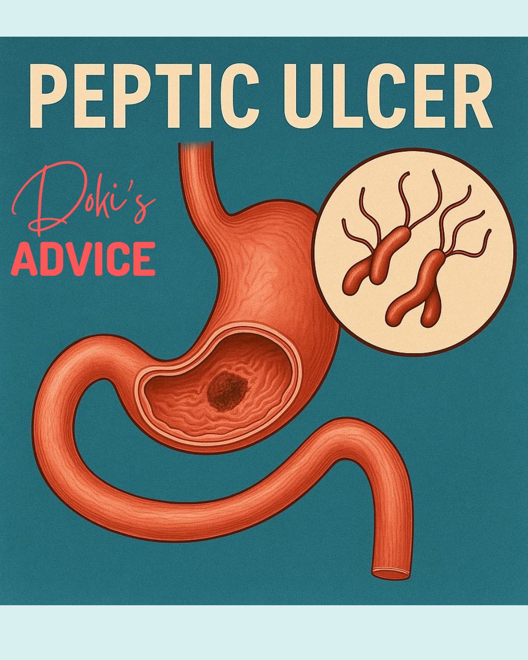
Moment...


Latest news

ALL ABOUT YOUR HEALTH!

We are determined to keep you regularly informed about our activities. Our Videos are meant to educate you more about your health! Karibu
Dokis-1Gall-Bladder-2.jpg
11/Jul/2024

Cholangitis: is an inflammation of the bile duct system. The bile duct system is found in the liver and is responsible for the transportation of bile from the liver, or to be precise, from the gall bladder, to the first part of the intestine, which is called the duodenum. Bile juice is made in the liver and stored in the gallbladder. In times of digestion, it’s then transported to the duodenum for the digestion of fats and small cholesterol.

Cholangitis is mostly caused by bacterial infections, and in other circumstances, it could be a chronic infection as the result of an autoimmune condition. An autoimmune condition is when the body identifies its own cells as foreign cells, hence attacking them.

Cholecystitis: this is an inflammation of the gallbladder either due to blockage of the bile duct system with a gallstone or bile sludge. Gallstones are small stones composed of cholesterol that usually form in the gallbladder. Gallstones are a common condition that affects nearly one in every 10 adults.

In medicine, we differentiate:

  1. Calculous cholecystitis: this is when the opening of the gall bladder, that is the cystic duct gets blocked by a gun stone or the bile sludge. This blockage then leads to a buildup of bile juice in the gall bladder, hence increasing the pressure inside, which can lead to inflammation.
  2. Acalculus cholecystitis: this is a guy bladder inflammation that is not caused by gallstones. This can be caused by severe diseases, operations, or traumas.
  3. Chronic cholecystitis: this is a result frequent acute cholecystitis which has been treated using conservative measures like giving antibiotics or healing spontaneously, hence leading to scarring of the gall bladder walls.

There are six major risk factors for the formation of gall stones, which include:

  • Being overweight
  • Women are more affected.
  • Fertile
  • Fair meaning white people or people with a fairly bright skin.
  • Age: around 40 years
  • When it occurs mostly in the family, the risk of other family members getting it is higher.

Cholecystolithiasis: simply refers to having gallstones in the gallbladder.

Choledocholithiasis: this is a term that refers to having gallstones in the bile duct system.

There are some underlying factors that can lead to more frequent building of gallstones. These factors include:

  • Disruption of the enterohepatic circulation: This can be caused by diseases such as Crohn’s disease or after recession of the terminal ileum which is the last part of the small intestine which can lead to bile acid loss syndrome.
  • Fasting, rapid weight loss, parenteral nutrition, or bariatric surgery
  • Haemolytic anaemia: a frequent destruction of red blood cells can lead to increased bilirubin, which can therefore lead to formation of pigment stones from the bilirubin. Bilirubin is a yellow substance that is made after the breakdown of old red blood cells.
  • Hyperparathyroidism: The condition that can lead to excessive calcium levels In the bloodstream, therefore contributing to the formation of gallstones out of calcium.
  • Caroli syndrome: this is a congenital disease that causes the deletion of the bile ducts in the liver intense can contribute to the formation of gallstones.
  • Gilbert syndrome: this is a genetic disorder of bilirubin metabolism that can increase the risk of gallstone formation.
  • cystic fibrosis: The condition that leads to a production of a thick mucus layer that can block the bile ducts hence leading to the formation of gallstones.

Let’s have a look at the signs and symptoms:

  • Nausea and vomiting
  • upper right abdominal pain
  • Painful cramps in the right upper abdominal region
  • Pain projection in the right shoulder
  • Fever
  • Jaundice
  • Itchiness

-Let look at how are these diseases diagnosed:

  • Your doctor will do a physical examination and take your medical history.
  • blood tests: Timeline whether the inflammation parameters are high and parameters of gallbladder obstruction, Liver and Pancreas.
  • Abdominal ultrasound: this is used to determine the structure of the gallbladder, to see if it is filled with gallstones, and to examine the anatomy of the liver.
  • endoscopic ultrasound: this can be used to detect small gallstones in the building system.
  • ERCP/MRCP: the former can be used for diagnostic and therapeutic purposes. The latter can only use for diagnostic purposes. This examination is used to showcase the biliary system. In cases of stenosis, the ERCP can be used for therapeutic purposes, for example, dilation of the biliary tracts, removal of gallstones and stenting. (This is a procedure where one can Insert a plastic or metal hollow tube to help relieve obstruction in areas of stenosis).
  • CT-Scan: mostly for purposes of better operation planning.
  • Abdominal X-Ray: done in suspicion of a hole or tear in the gall bladder.

Now let’s delve into how the above diseases are treated:

  • giving pain medications,
  • giving medication against cramping
  • Not eating or drinking to relieve the strain of the gallbladder.
  • Giving the patience fluids directly into the vein to prevent dehydration.
  • in cases of high inflammation parameters, giving antibiotics
  • Surgery for the removal of the gallbladder may be recommended after initial treatments to prevent acute cholecystitis and for the prevention of potential complications.
  • In other instances, surgery may not be recommendable, hence the importance of an ERCP, as stated in the diagnostics above.

So, you may ask yourself, are there ways to reduce the risk of getting cholecystitis?

  • Eating a healthy balance today while reducing the number of high cholesterol foods
  • Regular exercise reduces the risk of being overweight or obese.
  • avoiding Rapid weight loss diets

remember to always seek medical advice.


MeningitisMeningitis-2.jpg
07/Jun/2024

So, what do we understand by this?

Meningitis is an inflammation of the protective layer membrane that covers the brain and the spinal cord. Anyone can get meningitis, from small babies to older people, and it can be very severe, causing brain and spinal cord damage and long lasting disabilities.

Meningitis is caused by various agents, like bacteria (Neisseria meningitidis, pneumococcal bacteria, and bacteria causing tuberculosis), viruses such as HSV, chickenpox, and other harmless viruses. An infection caused by the HSV, chicken pox viruses, or bacterial meningitis is considered an absolute emergency that needs immediate treatment with antiviral or antibacterial drugs. Meningitis caused by bacteria can be treated by giving antibiotics, depending on the  bacteria that caused the meningitis in the first place, and it should be an antibiotic that can pass through the brain-brain barrier.

Before we delve into the topic, it is important to state that, meningitis can be prevented through vaccination. The meningitis vaccine can be given to children from the age of 12 months and all other age groups, especially those living within the meningitis belt. So, what exactly is the meningitis belt? These are the regions in Africa that are highly affected by meningitis, hence, a recommendation for the meningitis vaccine for kids of the age of 12 months as well as a pneumococcal vaccine.

Meningitis is spread by respiratory secretions through sneezing, coughing or kissing. In some cases, we do have meningitis carriers who can spread the infection yet they don’t suffer from it.

Children are mostly affected by bacterial variants, for example, the pneumococcal variant, which is also the leading cause of pneumonia.

What are some of the signs and symptoms of meningitis?

  • Headaches
  • high fever
  • nausea and vomiting
  • irritability to light
  • confusion
  • seizures
  • paralysis
  • Drowsiness or unresponsiveness
  • stiff neck
  • a rash that does not fade

The symptoms do not appear in any order and to some extent, some patients do not show any of the of the signs or symptoms stated above.

The incubation period for bacterial meningitis is around 2  to approximately 4 days but can also extend to 10 days. It is very important to recognise bacterial meningitis at an early stage and begin with antibiotic medication, as it is crucial because it can quickly end in death within hours or days.

The incubation period for viral meningitis is around 2 to 14 days. It mostly begins acutely, lasting a few hours to a few days. HSV or chickenpox virus caused meningitis, also needs early therapy with antiviral drugs. Other less harmless viruses that cause meningitis can heal by themselves after a couple of days to weeks. But since we cannot differentiate which viral infection or bacterial infection is causing meningitis, it’s always advisable to seek medical advice.

Let’s delve into the diagnosis of meningitis

  • clinical examination through your physician
  • inspection of your skin
  • examination of the meningitis sign
  • blood tests and microscopic examination of a blood stain
  • Lumbar puncture to collect cerebrospinal fluid for microscopic examination
  • in some cases, a CT-Scan can be necessary before the lumbar puncture. This is important in situations where symptoms such as unresponsiveness, loss of consciousness neurological symptoms that show suspicion of intracranial pressure signs are present.

So how do you treat a meningitis?

  • Early antibacterial treatments
  • supplementing the treatment with the antiviral treatments by suspicion of viral meningitis
  • cortisone in cases of intracranial pressure suspicion
  • supportive therapy such as
  • giving paracetamol or ibuprofen for pain or fever’s
  • Therapies for seizures in cases where epileptic seizures have been reported.
  • giving blood thinners in cases of septic thrombosis

Exposed people to those affected with bacterial meningitis are also given a prophylactic antibacterial medication to help reduce the Spread.

It is crucial to always begin an early antibacterial or antiviral therapy because if meningitis is not treated properly or early enough it could lead to severe long-term problems such as:

  • Hearing or vision losses
  • problems with memory and concentration
  • recurrent seizures afterwards
  • problems with coordination, movement, and balance
  • loss of limbs due to necessity of an amputation of affected limbs

Always seek medical advice and remember prevention is better than cure, so do get vaccinated as well as your children.


Cervical-cancer-doc.jpg
13/May/2024

Cancer of the cervix happens when normal cells in the cervix change into abnormal cells and grow out of control. Most people whose cervical cancer is found and treated early do very well. The cervix is the bottom part of the uterus, where it meets the vagina. The cervix is composed of two main types of cells. The outer layer of the cervix is covered with cells called squamous cells. Squamous cell carcinoma of the cervix is the name for a cancer that affects these cells. The cervix also includes glandular cells, which line the canal of the cervix that leads into the uterus. These cells can also become cancerous; when they do, they are called adenocarcinoma of the cervix. Although they arise from different types of cells, squamous cell carcinoma and adenocarcinoma of the cervix are treated similarly in the early stages.

Risk Factors

Most cervical cancers are caused by infection with a virus called human papillomavirus. HPV is spread by direct skin-to-skin contact, includi ng sexual intercourse, oral sex, anal sex, or any other contact involving the genital area (eg, hand-to-genital or mouth-to-genital contact). HPV infection can also cause a noncancerous condition called condyloma, which is caused by genital warts.

HPV infection is very common. Approximately 75 to 80 percent of sexually active adults will acquire a genital HPV infection before the age of 50. There are many types of HPV, and these affect different areas of the body. Most HPV infections are temporary because the body’s immune system effectively clears the infection.

Risk factors for cervical cancer include:

  • Smoking tobacco
  • Increasing number /constant change of sexual partners. The greater your number of sexual partners and the greater your partner’s number of sexual partners, the greater your chance of getting HPV.
  • Early sexual activity. Having sex at an early age increases your risk of HPV.
  • Bad sexual hygiene
  • Other sexually transmitted infections.
  • A weakened immune system, e.g HIV/AIDS
  • Genetic prädisposition
  • Exposure to miscarriage prevention medicine. If your parent took a medicine called diethylstilbestrol, also known as DES, while pregnant, your risk of cervical cancer might be increased. This medicine was used in the 1950s to prevent miscarriage. It’s linked to a type of cervical cancer called clear-cell adenocarcinoma.

Symptoms

Typically, cervical cancer develops slowly over several years. In some cases, the cancer does not cause any symptoms, while in others, it causes abnormal vaginal bleeding or discharge. This can include vaginal bleeding between menstrual periods, bleeding after sex, or bleeding after menopause. This bleeding may be spotting or heavy bleeding.

Other symptoms may include:

  • Menstrual bleeding that is heavier and lasts longer than usual.
  • Watery, bloody vaginal discharge that may be heavy and have a foul odor.
  • Pelvic pain or pain during intercourse.

Diagnosis

Pap testing and human papillomavirus (HPV) testing are commonly used together to screen for cervical cancer. If a Pap test shows abnormal cells, further testing is essential, as treatment of abnormal cells of the cervix can prevent cervical cancer. A biopsy of the cervix involves removing a small piece of tissue from the cervix. The biopsy is performed during an office visit using a procedure called colposcopy. The colposcope, which is similar to a large magnifying lens, magnifies the view of the cervix. This allows the clinician to better see the location, extent, and degree of cervical abnormalities that may not be visible with the naked eye alone.

The tissue obtained during the biopsy is examined with a microscope to see if cervical cancer cells are present. In some cases, more of the cervix will need to be removed for biopsy; this is done through a procedure called cervical conization or a cone biopsy. This can be done either in the operating room by a surgeon using a scalpel or in the office using a loop electrosurgical excision procedure (LEEP). LEEP is performed with a device that uses an electric current to remove a piece of the cervix. You will usually get the results of the biopsy one to two weeks after the biopsy is done.

CT-scan or MRI of the Pelvis Region: to determine spreading of the tumor to other organs or regions of the body.

Treatment

Cervical cancer can be treated in different ways:

  • Surgery: Some cases of cervical cancer are treated with surgery to remove the cancer. Different types of surgery can involve:
  • Removing the cervix, uterus, and upper part of the vagina is called a “radical hysterectomy.”
  • Removing all or part of the cervix but leaving the uterus in place—this surgery is done only in special situations.
  • Radiation therapy: Radiation kills cancer cells.
  • Chemotherapy: Chemotherapy is the medical term for medicines that kill cancer cells or stop them from growing. People with cervical cancer usually receive chemotherapy at the same time as radiation therapy.

Prevention

Cervical cancer can be prevented through:

  • HPV Vaccination.
  • Routine Pap tests.
  • Practicing safe sex.
  • Proper Hygiene
  • Avoid cigarette smoking.

Dokis-Pneumonia-2-1200x1200.jpg
04/Apr/2024

Pneumonia

Pneumonia is an inflammation of the lungs that is mostly caused by bacterial infections but can also be caused by viral infections. It is the most common reason for deaths in developing and in industrial nations.

In Kenya, pneumonia is responsible for approximately one in five children’s deaths. It claims approximately 8,000 to 10,000 children’s lives annually.

Pneumonia takes about two to four weeks before healing. For small children and older people people with lung and heart conditions, the infection will take longer and even probably require hospitalization due to the severity of their conditions.

As mentioned earlier, pneumonia is caused by vectors such as bacteria and viruses, and in patients with a reduced immune system, pneumonia can also be caused by fungi.

Some of the factors that increase susceptibility to pneumonia infections include:

  • age factors such as young babies and older people
  • seasonal factors like rainy and cold seasons
  • Prior medical history such as asthma in COPD (chronic obstructive pulmonary disease),

Pneumonia can be categorized into:

  • community-acquired pneumonia: the most common bacteria are pneumococcal, and Haemophilus influenza, others include: Chlamydophila pneumococcal, Mycoplasma pneumococcal, Legionella, Staphylococcus aureus, and Mycobacterium tuberculosis.

-Viruses such as: Influenza, RSV, and Corona Virus

  • Hospital-acquired pneumonia: the most common bacteria for these are Pseudomonas aeruginosa and Staphylococcal.
  • Pneumonia on patients with a reduced immune system.

mostly caused by minor bacteria and fungi

  • Neonatal Pneumonia

Let’s look at signs and symptoms of pneumonia:

  • Fever
  • Coughing up yellow or green mucus
  • Chest pains and pains while breathing
  • Aching of the body, joints and headaches
  • Exhaustion
  • Loss of appetite
  • Wheezing while breathing
  • Confusion and hallucinations in older patients or due to certain viral infections like influenza

Now that we know the signs and symptoms to look out for, let’s dive into how we diagnose pneumonia:

  • Clerking: this is when the doctor asks general questions about your cough, including when the symptoms started and any prior medical history
  • the doctor checking for signs like strenuous breathing and blueness of the lips and tongue, this is due to less oxygen intake in the lungs.
  • The doctor will do a physical examination using his stethoscope to determine the different sounds of the lungs while breathing
  • Blood tests: this helps to determine between a viral and a bacterial infection. In the case of a bacterial infection, the white blood cells and inflammation parameters are mostly high. In the case of a viral infection, the white blood cells and inflammation parameters will be normal or just very slightly elevated. In other cases, one could have both a viral and a bacterial infection, this is what you would call a superinfection in medical terms.
  • X-ray: this helps to determine where exactly the pneumonia is located; for example, it could be on both sides of the lungs or just on one side.

Now that we know the different diagnostic steps, let’s have a look at how he treats pneumonia.

  • Bacterial infections are always treated with antibiotics.
  • Viral infections do not require antibiotics. Mostly, this kind of pneumonia just requires anti-inflammatory treatment with medications such as Ibuprofen, and paracetamol, and enough fluid intake.
  • In case of a superinfection with a bacterium and a virus, antibacterial therapy will be required.
  • In case of a fungal infection, antifungal therapy is required.

Please note that not all kinds of pneumonia require antibiotics, hence the need to draw blood to differentiate between a bacterial, viral, or fungal infection. This is crucial to avoid the high rise of resistance in antibiotic therapies.

We do say prevention is better than cure, so how can we reduce the frequency in which pneumonia occurs in our general population? The main method of pneumonia prevention, especially in small children and older patients is vaccination.

-small babies and children can be vaccinated against Haemophilus and pneumococcal disease at the age of two months. Older patients as well as patients with a reduced immune system can also prevent the severity of pneumonia by getting vaccines against the named bacteria above.

For more medical gems, keep at Doki’s Advice.


Dokis-colorectal-2.jpg
17/Mar/2024


This is a malignant tumour found in the lining of the large intestines up to the rectum.

The large intestine is known for its role in the digestive system to absorb water and remove waste from the food we eat.

Colon and rectal cancer mostly occur at older age, but by a particular number of people who have certain gene mutations, this cancer can occur at an early age, for example, at 30 without them realising it.

Colorectal cancer is the third most common cancer in the world and the second leading cause of death globally.

According to the data required from Globocan, there were an estimate of 1,354 new cases and 937 deaths from colorectal cancer in Kenya in 2018. Other data sources have noticed that there is a rise in colorectal cancer cases in Kenyan rural areas and that it is the fifth most common cancer in sub-Saharan Africa.

Let’s delve into factors that increase the risk of colorectal cancer:

  • Genetic Factors:
    • Such as familial disposition
    • Familial polyposis syndrome: Families with this syndrome tend to have the growth of many polyps along the large intestines at a very young age. These polyps can with time, go through genetic mutations, causing them to be cancerous and hence leading to early-stage colorectal cancer. To note, with this syndrome, the growth of polyps can also occur in any part of the intestines.
    • Hereditary nonpolyposis colorectal cancer syndrome: This is also another genetic disease that increases the risk of colorectal cancer.
  • Eating habits and lifestyle:
    • Smoking and excessive drinking of alcohol.
    • Obesity
    • Low fibre or high fat diet
    • Red meat or processed meat and foods

-Some protective factors would be:

  • Exercise
  • Eating foods rich in fibre, vegetables, and less meat
  • Maintaining healthy weight

Now that we know the risks and protective factors, let’s have a look at the signs and symptoms of colorectal cancer:

  • Most of the time, there are no characteristic early symptoms
  • A constant change of bowel movements from Obstipation and diarrhoea
  • Rectal bleeding: this can either be visible blood or non-visible, meaning it can only be detected by doing a faecal occult blood test.
  • Persistent abdominal discomforts such as cramps passing gas or pain
  • Unexplained weight loss within a short period of time
  • Feeling like the bowl isn’t completely empty even after passing stool.
  • Fatigue and weakness
  • Unexplained Anaemia


So how do we diagnose colorectal cancer?

  • A basic diagnostic is performing a digital rectal examination from your doctor. This is used to detect palpable Tumours just near or around the rectal and anal regions.
  • Complete colonoscopy with biopsy: This is an examination of the whole large intestine with the possibility of taking biopsy samples to pathologically test the sample for tumours type.
  • Rectoscopy for rectal cancer diagnosis.
  • CT-Scan for staging purposes: is the tumour metastasized? if yes to what organs.
  • Lab tests for tumour markers such as CEA as a control value especially while doing chemotherapy and years after remission.
  • MRI-Scan: mostly in regard of rectal cancer.

After a proper diagnosis of colorectal cancer the next step would be to discuss with your oncologist and surgeon what the individual therapy plan will look like. This is determined by factors like

  • how physically fit is the patient prior to therapy,
  • is the patient eligible for surgery or not,
  • size of the tumour
  • is the tumour only at one spot or several spots in the bowel?
  • does the tumour have certain genetic changes?
  • Is prior Radio-chemotherapy necessary or not.

-Let have a look at the different therapy options

  • Surgery: This is the option for tumours that are just locally on one spot or multiple tumours localised on the same segment of the large intestine. Some tumours could have spread to the liver but could still be operable when the metastasis is either singular or multiple but localised in the same area. Such factors would have to be considered by your surgeon.
  • Chemotherapy: Some Tumours could be so large that prior chemotherapy is required to shrink them to an operable size.
  • Radiotherapy: This is mostly done on rectal cancers since colon cancers are insensitive to radiation.

Which therapy regime each patient needs depends on some of the factors noted above. Some may need a Stoma bag after an operation. This will also be discussed with you during the planning stage for the therapy needed.

As always, seek medical advice and keep it Doki’s Advice for more info gems.


Dokis-vertigo-1200x1200.jpg
04/Mar/2024

It does not in totality equate to dizziness but is a form of dizziness accompanied by a whirling, spinning sensation with a lack of postural stability or balance. Can be accompanied with Nausea and Vomiting

Vertigo can be a disease on its own, but it can also be a symptom of a chronic or acute condition.

For a better understanding, let’s remotely understand how balancing works. Balancing involves a complicated connection between the brain, inner ears, eyes, spine, and muscles. Disturbances, injuries, and infections of the above-mentioned can result in dizziness or vertigo.

Causes range from benevolent to life-threatening conditions.


Let’s start with the common ones:

Side effects of Medications such as antibiotics, chemotherapy, etc.

* Specific forms of migraines, like vestibular migraines.

*Motion Sickness.

peripheral Vertigo involving the inner ear (vestibular organ)


Today we focus on peripheral vertigo as a result of malfunctioning of the inner ear.

It’s important to note that the inner ear, being one of the smallest organs, has a complex ib structure with very many microscopic structures coordinating earing and balancing. We will spare you the details of how it works.

Peripheral vertigos include:

  1. Benign paroxysmal positional Vertigo.

The most common vestibular disorders.

Vertigo or dizziness with acute onsets may last a few seconds to a few minutes.

The vertigo manifests itself with a change in body posture: turning in bed, movement of the head.

Trigger in layman’s tongue: irritation of the inner ear due to loose microscopic fragments detaching themselves from their fixation and circulating within the microscopic inner channels of the ear.

The diagnosis is clinical.

Therapy is the repositioning of detached fragments with physio—therapeutic special vertigo training.


2.Neuropathia Vestibularis.

Patients present with severe vertigo, mostly associated with nausea and vomiting and conspicuous loss of balance.

It is believed to be associated with an inflammation of the inner ear.

The diagnosis is also clinical after a thorough examination to rule out possible central causes.

Patients profit from steroids such as prednisolone and other symptomatic medications like vomex.

3.Meniere Disease.

Presents with a triad of tinnitus, loss of hearing, and severe vertigo.


Red flags of acute dizziness or vertigo:

In acute onsets, it is important for a clinician to note that most Vertigo’s are harmless. Underlying disorders may be overseen.

These include strokes of the brainstem, tumors, or even cardiac conditions.

It is important to rule out these conditions.

Using MRI scans. Long-term ECGs.

The most important thing is to always do a thorough clinical examination and a well-sourced patient’s history. When in doubt as a clinician, better do more tests than ignore your instincts.


Lastly, as a patient, before presenting yourself with dizziness, ask yourself simple questions like:

Did I hydrate well? Is my blood sugar okay?

And, as always, seek medical advice for better treatment.


Dokis-Prostate-cancer-1200x1200.jpg
04/Mar/2024

Prostate cancer is one of the most common types of cancer. Prostate cancer occurs in the prostate, which is a wall-nut-shaped gland in males. The function of the prostate is to produce seminal fluid that transports and nourishes sperm. Many prostate cancers grow slowly and are confined to the prostate gland, where they may not cause serious harm. However, while some types of prostate cancer grow slowly and may need minimal or even no treatment, other types are aggressive and can spread quickly. Prostate cancer, when detected early, has a better chance of being successful.

What are the risk factors for prostate cancer?

It’s not clear what causes prostate cancer. However, the following risk factors may play a role:

  • Race: For reasons not yet determined, black people have a greater risk of prostate cancer than do people of other races. In black people, prostate cancer is also more likely to be aggressive or advanced.
  • Obesity: People who are obese may have a higher risk of prostate cancer compared with people considered to have a healthy weight, though studies have had mixed results. In obese people, the cancer is more likely to be aggressive and more likely to return after initial treatment.
  • Older age: Your risk of prostate cancer increases as you age. It’s most common after age 50.
  • Family history: If a blood relative, such as a parent, sibling, or child, has been diagnosed with prostate cancer, your risk may be increased. Also, if you have a family history of genes that increase the risk of breast cancer (BRCA1 or BRCA2) or a very strong family history of breast cancer, your risk of prostate cancer may be higher.


What are the symptoms of prostate cancer?

Prostate cancer may show no signs in its early stages.  When advanced, it may cause the following symptoms:

  • Trouble urinating
  • Decreased force in the stream of urine
  • Blood in the urine
  • Blood in the semen
  • Bone pain
  • Losing weight without trying
  • Erectile dysfunction

What are the complications of prostate cancer?

  • Metastasis: Spread to nearby organs, including the bones and bladder.
  • Urinary Incontinence: Inability to control passage of urine
  • Erectile dysfunction


How is prostate cancer diagnosed?

  • PSA Testing: This is the most commonly used and most valuable test for early detection of prostate cancer. The likelihood of prostate cancer increases with more elevated PSA values.
  • Age-specific ranges include
  • 40 – 49 years (0 to 2.5ng/mL)
  • 50–59 years (0 – 3.5 ng/mL)
  • 60 – 69 years (0-4.5 ng/mL)
  • 70–79 years (0 – 6.5bng/mL)
  • Digital Rectal Examination (DRE): This involves the insertion of the digit finger into the rectum to assess for prostate enlargement. DRE may detect psotate nodules, induration, or asymemetry that can occur with prostate cancer.
  • Transurethral ultrasound is often used to evaluate abnormalities detected on DRE.
  • Prostate Biopsy: Depending on the findings of DRE, PSA, and prostate ultrasound, a biopsy of the prostate may be recommended. Biopsy is done to obtain tissues for histologic diagnosis. This is the definitive diagnosis for prostate cancer.


How is prostate cancer treated?

  • Active Surveillance: This involves routine tests to check whether the cancer is starting to grow more quickly. If so, active treatment should be initiated.
  • Surgery: This involves the removal of the prostate gland through surgery.
  • Radiation Therapy: Radiation kills cancer cells. Radiation can be given from a machine that moves around the body or from a direct source to the prostate gland.
  • Hormone Therapy: The male hormones are responsible for the growth of the prostate gland. Hormone therapy reduces the levels of the hormones and thus shrinks the cancer. Hormone therapy can be done through the removal of testicles or through medications.
  • Chemotherapy: Advanced prostate cancer requires chemotherapy for complete treatment.

Remember to always seek medical advice from your practitioner.


30/Jan/2024

In Kenya, cancer is the 3rd leading cause of death, accounting for approximately 7% of the deaths per year, after infectious diseases and cardiovascular diseases.

Generally, it’s difficult to get accurate data due to circumstances such as:

Low collection accuracy and underdiagnosis. A huge number of cancer patients could go undiagnosed, especially if they lack adequate funds for diagnostics and treatment, hence avoiding going to hospital.

Most of the data collected comes from Nairobi and other urbanised settings.

Most leading cancers in Kenya are:

  • Breast cancer: 34 cases per 100,000 people
  • Cervical cancer: 25 per 100,000 people
  • Prostate cancer: 17 cases per 100,000 people
  • Oesophageal cancer (food gut): 9 per 100,000 people

Approximately 75% of cancer cases are diagnosed in late stages due to factors named above as well as lack of awareness.

So, let’s dive into lung cancer as well as other types of cancers we will handle in coming articles.

Lung cancer is a malignant tumour growth that can occur in either side of the lungs or both. This can either be the main tumour or a metastasis (cancer spreading) of other types of cancers of the prostate, breast, head, neck, kidney, and womb, just to mention a few.

The downside of most cancers is that they have no initial symptoms that one could watch out for, in order to push one to go for an early check-up.

Nonetheless, lung cancer patients could experience common symptoms such as:

  • Persistent dry coughing
  • Coughing up blood
  • Breathlessness or difficulties breathing.
  • Unexplained loss of weight: particularly a huge amount of weight in within a short period of time
  • Signs of anaemia: Loss of breath, getting tired quickly, pale face, heart palpitations
  • Unexpected loss of appetite
  • Pain while coughing or breathing.
  • Problems swallowing
  • Hoarseness of the voice
  • Intermittent night fevers, sweating at night.

There are two main types of lung cancer:

  • Non-small-cell lung cancer: this is the most common form, accounting for about 85% of most cases. It grows slowly before getting to the size in which it could start causing symptoms.
  • Small-cell lung cancer accounts for about 15% or less of lungs cancers. It has a very fast growth rate hence could lead to quick deterioration of a patient’s symptoms due to early metastases (spreading).

The type of lung cancer a patient has determines the kind of therapy they will require.

The age at which most people are affected is around the age of 40 and above. Rarely does it occur to younger patients.

The most common cause of lung cancer, as we all know, is of course, smoking, which accounts for approximately 70% of the cases. This does not mean that nonsmokers cannot develop lung cancer.

Other causes of lung cancer among nonsmokers are:

  • Long exposition to asbestos and radon exposure (a few studies have found high exposure to radon in the coastal region, especially in Taita/Taveta region and around Nairobi Kenyatta University).
  • Exposure to Industrial gases and vehicle exhaust gases
  • Exposure to polycyclic aromatic hydrocarbons: like cooking indoors with wood, there is a constant inhalation of the gases produced. This has also been seen to cause food-gut cancers.
  • Exposure to some metals like: Arsen, chromium or Nickel

Let’s see how lung cancer can be diagnosed:

  • X-Ray: though with this method, it’s rare to be able to see the tumours
  • CT-Scan: This is the ideal method of diagnosis to determine the areas of metastasis, like the bone and brain.
  • Getting a sample of the growth and sending it in for pathological testing to prove if its malignant or not.
  • One can also do lab tests for markers in the blood.

– NSE: Mostly high in small-cell lung cancers

–  LDH: generally, suspiciously high in most cancer patients, regardless of the type of cancer

– CYFRA-21: mostly determined to check therapy responsiveness.

Now that we know what methods we use to detect, lets dive into diff therapy regimes. These are also determined by how big the tumour is, how many of them are there, either on one side of the lung or both, and how far it has spread to other organs.

  • Curative operative resection: This is mostly specific for small tumours that haven’t metastasized yet and are locally on one side of the lung and easily operatively reachable.
  • Combination of operative resection and chemotherapy: This is also ideal for tumours that are relatively big but still operatively reachable for resection after going through chemotherapy with the intent to reduce the size of the tumour. Hence, this can also be done with an aim of curing.
  • Combination of an operation, chemotherapy, and radiotherapy: This is done when the tumours are, for example, on both sides of the lungs and/or metastasized to the liver. One could either begin with chemotherapy to reduce the size of the tumours followed by an operative resection of the tumours or, in the end, do radiation therapy to ensure that all tumour masses that couldn’t be removed during the operation are also treated. Otherwise, one can do simultaneous chemotherapy and radiation therapy and at the end, do the operative resection.
  • Palliative chemotherapy: this is does when the tumour isn’t operatively resectable and due to the presence of too many metastases. This means this can be done with the aim of just trying to shrink the tumour, to reduce the symptoms and not for the purpose of curing it.
  • Radiation of the brain: This is done in cases of brain metastases with or without the option of an operation.

-Which therapy regime is appropriate for each patient is dependent on the stage of the cancer at the time of diagnosis, the patient’s capability to withstand a particular therapy regime, and their general health situation.

-Always seek medical advice from a professional oncologist, surgeon, and oncology-radiologist because the treatment of a lung cancer patient is a multidisciplinary therapy concept.

-Other than that, avoid smoking. If you are already a smoker, do your best to stop smoking or do smoking cessation therapy.

And as always, for more medical gems, keep it Doki’s Advice.


diabetes-Type-1-2.png
02/Nov/2023

-This is one of the most common conditions in the world currently affecting the whole world in large numbers.

-3% of adults in Kenya are affected by diabetes, but in this number, we haven’t considered the large number of children and adults who are undiagnosed.

-So, I know most of have heard the word Diabetes and have an idea that it’s a condition whereby blood sugar levels are too high. I want us to dive deep into this disease to better understand it and how much damage long term high blood sugar levels affects those with diabetes.

-Before we get into that it’s good to clarify that there are two main types of diabetes known by the general public, type 1 and 2 but there exit two other types. Let’s have a look at all 4:

  1. Type 1 Diabetes: This is where the pancreas (organ of the body responsible for producing insulin, glucagon, and other digestive hormones) is incapable of producing insulin (the hormone responsible for reducing or rather regulating high blood sugar levels).
  2. Type 2 Diabetes: This is where the pancreas can’t produce enough insulin or in other cases where the body cells are not able to take up insulin as required, what we clinically sometimes say insulin insensitivity of some body cells.
  3. Type 3 Diabetes: Now this one I bet so many of us might not know exits but this is when the pancreas is damaged hence creating its inability to produce enough insulin. This type can happen after:
  • an acute or a chronic phase of pancreatitis (This is an inflammation of the pancreas mostly caused by excessive consumption of alcohol or due to bile stone obstruction of the pancreatic duct). Now don’t worry we’ll talk about pancreatitis in a topic of its own.
  • pancreatic cancer
  • cystic fibrosis: This is a genetic condition that cause damage of the lungs, digestive organs like the pancreas and other organs in the body.
  • Haemochromatosis: This is also a genetic inherited condition which leads to high iron levels accumulation in the body which in turn can lead to the damage of organs such as the liver, pancreas, and the heart.
  1. Gestational diabetes also Type 4: This occurs during some pregnancies and tends to disappear after birth, but in some women, it may persist afterward then as type 2.


-Now that we know the various types of diabetes let’s start from the top.

Type 1 Diabetes

-This type of diabetes can develop from a young age mostly to around the age of 25 to 30 years. The downside of it is that it can also start at any age but isn’t as common.

-As earlier stated, the pancreas gradually reduces the production of insulin until it eventually doesn’t produce any at all. The amount of blood sugar then increases over time, which can cause serious damage to organs like the kidneys, blood vessels, eyes, and nerves.

Cause oy type 1 diabetes:

  • The main cause is due to an autoimmune condition: this means one’s body mistakenly identifies pancreas cells are harmful and attacks and destroys them. Hence its function, production of insulin that enables blood sugar to be taken up by the needed cells such as muscles can’t be executed.
  • It’s also associated with other autoimmune conditions like: Type-A-Gastritis, celiac disease (gluten intolerance) or Addison’s disease (inadequate production of steroid hormones due to adrenal insufficiency).

Symptoms of type 1 Diabetes

  1. Excessive thirst
  2. Urinating frequently than usual and mostly at night
  3. Being constantly tired
  4. Weight loss
  5. Loss of muscle
  6. Blurred vision
  7. Having frequent urinary tract infections: mostly yeast infections: due to higher sugar concentration in urine.
  8. In later stages when the blood sugar levels are in extreme highs:

– deep breathing,

– nausea and vomiting

-a fruity smelling breath

-falling into a coma

Diagnosis of type 1 diabetes:

  • Urine tests: this is used to test for something called ketones (end-product when the body breaks down fats to give the body energy) and glucose in urine.
  • Blood tests:

-HbA1c test- this is a test checking the level of glycated haemoglobin. This value can also be used to control how good one’s diabetes is being managed.

-Determining of the C-peptid: This is a part of an insulin molecule. In type 1 diabetes patients its missing since they can’t produce insulin at all.

  • Testing the amount of blood sugar levels after a certain period of fasting, mostly after 8 hours of fasting.
  • Specific test to determine certain antibodies in type 1 diabetes.

Intervention/Treatment:

-The basic rule of treatment of diabetes type 1 is that all patients need an insulin therapy. If you remember, we said earlier that a type 1 diabetes patient doesn’t produce any insulin. This means these patients require insulin to enable them to regulate and propel the uptake of blood sugar in the required cells.

-Now how much insulin each patient requires depends on different criteria’s such as: Eating habits, body activity, and severity of insulin resistance in the body.


Insulin therapy schemes:

Conventional insulin therapy:

-This is giving fixed insulin doses at specific times of the day e.g., in the morning and evening. This would also mean the patients need to eat at specific times and specific amount of bread units, since the insulin doses are particularly fixed depending on this cycle.

-So, what is a bread unit (BU) or as some may call it a carbohydrate unit (CU)? This is used to calculate the amount of carbohydrates contained in food. One BU is equal to approximately 10 to 12g of carbohydrates.

-Example of how this works: Patient A with type 1 diabetes applies the insulin at 7am in the morning and 7 pm in the night. 2/3 of the insulin doses is therefore injected at 7am and 1/3 of the doses at 7pm. The patient is then required to make sure that they have their meals at specific times and specific amounts of bread units e.g., breakfast at 7:30am lunch at 13pm and dinner at 7:30pm.

Intensified insulin therapy:

-In this therapy regime the intention is to imitate the natural blood sugar management in the body.

-How do we go about this?

-patients inject themselves a long-acting basal insulin (e.g., insulin glargine, insulin Levemir): this can be done either once a day or twice a day

-Patients then inject a short-acting insulin depending on the meals they take and blood sugar levels at the time of application. (e.g., insulin lispro, insulin apart, insulin glulisine or normalinsulin)

Insulin pump therapy:

-This type of therapy is where the patients get an external pump on the body that continuously injects insulin depending on the blood sugar level.

-This therapy regime is mostly applicable for children as they are not capable of controlling and doing the self-application of insulin or also for adults whose blood sugar management isn’t satisfactory.

-The downside about this therapy is that it’s important for the patients to frequently check their blood sugar levels to avoid hypoglycaemia which can lead to a diabetes coma and if not treated early could lead to death.

-Please note for diabetes treatment: always consult your doctor and do regular check-ups to check if your diabetes management is under control, hence helping reduce diabetes complications. We’ll talk about these complications after looking at all the other types of diabetes.


Type 2 Diabetes

As explained on our previous article, in type 2 diabetes, the body is either incapable of producing enough insulin to breakdown blood sugar through sugar uptake in the necessary cells like the liver, muscles, kidney, fatty cells or the brain cells, or the insulin sensitivity is so low that blood sugar uptake in the named organs is not guaranteed.

In most cases type 2 diabetes Is associated with the metabolic syndrome.

-What is this syndrome you may ask? It’s a term used to describe a combination of diabetes, high blood pressure, abnormal cholesterol and obesity that together increase the risk of heart diseases, stroke and other conditions affecting the blood vessels.

Symptoms of a metabolic syndrome:

  • Being overweight, mostly having a pear shape or apple shape around your waist.
  • High levels of the ‘bad type of fats’ in the body (medically called LDL cholesterol) and less of the ‘good type of fats’ (medically called HDL cholesterol).
  • High blood pressure of over 140/90 and higher
  • Diabetes

Complications of a metabolic syndrome can include:

  • Cardiovascular diseases such as:
  • High blood pressure
  • Diabetes
  • NASH (Non-alcoholic fatty liver disease) Check out our Liver cirrhosis articles to find out what this is.
  • Tumours

Symptoms of type 2 Diabetes: just as type 1

  1. Excessive thirst
  2. Urinating frequently than usual and mostly at night
  3. Being constantly tired
  4. Weight loss
  5. Loss of muscle
  6. Blurred vision
  7. Having frequent urinary tract infections: mostly yeast infections: due to higher sugar concentration in urine.
  8. In later stages when the blood sugar levels are in extreme highs:

– deep breathing,

– nausea and vomiting

-a fruity smelling breath

-falling into a coma

Diagnosis of type 2 diabetes:

  • Urine tests: this is used to test for something called ketones (end-product when the body breaks down fats to give the body energy) and glucose in urine.
  • Blood tests:

-HbA1c test- this is a test checking the level of glycated haemoglobin. This value can also be used to control how good one’s diabetes is being managed.

– When your blood is drawn by your doctor the HbA1c is the  value they determine to see if it lies within the normal margin or  above (normal values would be between 4.5% and 5.7%…..For values above 6.5% one would the give the diagnosis: Diabetes). Now disclaimer this value may also slightly differ depending on the parameters each lab uses.

-Determining of the C-peptid: This is a part of an insulin molecule. In type 1 diabetes patients its missing since they can’t produce insulin at all.

  • Testing the amount of blood sugar levels after a certain period of fasting, mostly after 8 hours of fasting.


Interventions/Treatment:

-Now here there is one slight difference to type 1..and that is one can start with oral medication unlike type one where insulin application is non-negotiable.

-Now since we live in a modern society therapy is a bit modified to fit each patient specifically. This kind of modified type of therapy takes into consideration a patients Life expectancy, other ailments, other medications that they take, risk of someone having hypoglycaemia and so on.

-There are different type of antidiabetic medications for example:

  1. Change of Lifestyle: through better eating habits and exercise,
  2. Metformin: Now this is one of the most prescribed antidiabetic drugs. It can be used as a mono therapy. This means just prescribed alone especially if the patients long term blood sugar isn’t too high.

-Now at this point I know your asking what is a long term blood sugar value right?. This is the  Hb1Ac value measured from your red blood cells as described above and it can also be used to determine how good the given therapy inclusive of eating habits have been working for at least the last 3 Months. Why 3 Months you ask? Because red blood cells have a 3 Month life expectancy before they are replaced with new cells.

  1. Combination Therapy: example

– Metfromin + Dapaglifozin /Empaglifozin

– Metformin  + Dulaglutid/Liraglutid

– Metformin + Glibenclamid

-Metformin + any one of the above + insulin

Disclaimer: You should always consult a medical doctor before taking any medication for advice. This because some of these medications have different side effects and could be inappropriate for one patient but appropriate for another.

  1. Insulin therapy as discussed in our previous article in type 1 diabetes.


Some side effects from some of these therapy regimes include:

  • Weight loss: mostly this is usually a desired effect.
  • Headaches
  • Stomach issues: Bloating, nausea, stomach pains, too much gas etc.
  • Inflammation of the pancreas (Organ responsible for production of insulin and other digestive enzymes)
  • Hypoglycaemia: This is one of the most dangerous side effects because it could lead to a coma and death in a very short time, hence why its critical that one consults a medical doctor and be taught what to look out for during a hypoglycaemia.

-Signs such as:

  1. Dizziness →Coma
  2. uneasiness
  3. sweating
  4. heart palpitations (Like your heart beating very fast that its uncomfortable)
  5. nausea and vomiting
  6. Confusion
  7. Headaches
  8. Difficulties talking
  9. Seizures
  10. Loss of concentration

Want to find out more on diabetes? Keep it at Doki’s Advice and remeber to always seek medical advice before any treatment and when you notice any of the symptoms above.


Gastrointestinal-Bleeding-1.png
08/Oct/2023

Upper gastrointestinal Bleeding (GI-Bleeding)

-This is bleeding that occurs in the upper gut, stomach and/ or the duodenum (this is the first part of the small intestine).

-What do you have to look out for if you suspect that you are having upper GI-bleeding?

Here are some signs:

  • Most obvious one is if you vomit blood: this can be red or dark brown resembling the colour of coffee.
  • Black, tarry stool: now here we must caution, because someone taking iron medication or medication containing iron tend to also have black stool. For such people this wouldn’t be a sign of bleeding.
  • Dizziness and/ or Fainting: this is due to loss of blood.
  • Weakness and getting tired too quickly: also due to blood loss.
  • Heartburn: now warning this is a very general symptom that occurs to many of us. That means this alone doesn’t ascertain that one has an upper GI-Bleeding.
  • Nausea: also, a very general symptom, but mostly occurs in combination with vomiting blood.
  • Symptoms of shock

-What should we understand by this? This is when one loses a large amount of blood that the body suddenly goes into a shock mode:

-What to look out for when in shock?

  • Sudden drop of blood pressure
  • A very fast pulse rate
  • Not urinating at all or very minimal amount of urine in the cause of the day: this is caused by reduced amount of blood flowing to the kidneys, hence causing an acute kidney failure.
  • Unconsciousness

-If this happens seek medical attention ASAP!

-Now that we know what signs to look out for, lets dive into the various causes of upper GI-Bleeding.

  • Peptic ulcers  ̴40%: this are sores on the deeper lining of the intestinal tract. These sores can occur in any part of the intestine. When the deeper lining is destructed by too much stomach acidity or infections or medication, it can be so severe to the point of damaging blood vessels running underneath the intestinal lining hence causing GI-Bleeding.
  • Causes: H-pylori infections, anti-inflammatory drugs (Ibuprofen)
  • Gastric erosions ̴20%: this are sore on the upper lining of the stomach. This can occur due to alcoholism, frequent us of medication such as ibuprofen,

-Signs of gastric Ulcers: heartburn, indigestion, or reflux.

  • Varices bleeding  ̴15%: these are swollen or large blood vessels that develop in the gut and the stomach mostly due to a chronic liver disease such as Liver cirrhosis. They develop when the normal blood flow to the liver is blocked either due to scaring or a blood clot, hence causing blood to flow in smaller blood vessels, that in normal circumstances, can only transport blood in small amounts. When the blood in these vessels increases, there stands a risk of them busting open, hence upper GI-Bleeding.

-Stomach acidity can increase due to factors such as: bacterial infections or medication like Ibuprofen taken for a longer period without taking PPI’s (PPI are drugs meant to reduce the acidity produced by special stomach cells).

  • Gut and stomach tumours: When the stomach lining is weakened or a penetration of the tumour through a blood vessel.
  • Mallory Weiss tear: This is a tear that occurs along the gut due to prolonged vomiting or coughing. The Lesions caused can bleed so much that an intervention would be necessary to prevent excessive blood loss.

Prevention and Treatment of upper GI-Bleeding

Avoiding excessive consumption of alcohol

Avoid excessive smoking.

Early and thorough treatment of bacterial infections caused by H-Pylori

By prolonged use of anti-inflammatory drugs supplementary use of PPI’s (These are medication to help reduce stomach acidity).

In cases of bleeding, seeking immediate medical attention with interventions such es endoscopic haemostasis


Lower Gastrointestinal Bleeding (GI-Bleeding)

Now let’s jump into the lower part of the GI-Tract and look at the signs and symptoms, evaluate causes, and look at the interventions.

Lower GI-Bleeding is occurring in the lower gut. Lower gut is considered to start from lower small intestine and the whole large intestine till the rectum.

Let’s have a look at some signs and symptoms:

General symptoms:

  • Pale Skin due to blood loss
  • Feeling weak or getting tired quicker than normal, even after doing work that requires less amount of energy.
  • Heart palpitations
  • Fainting
  • Dizziness

Specific Symptoms for lower GI-Bleeding:

  • Red blood mixed with stool or layering on stool.
  • Uncommon: black stool when the bleeding is located at the lower small intestine (medical term Ileum or caecum).
  • Increased stool frequency: blood sometimes acts as a laxative in the lower GI-tract.

Now that we know what to look out for let’s see what causes bleeding of the lower GI-Tract

  1. Haemorrhoids: these can be large or small lumps of blood vessels located on one’s rectum that tend to cause bleeding especially due to hard stool like during obstipation. Many people suffer from this, so there’s no need to feel ashamed. Signs are:
  • Bright red blood layered on stool.
  • Itchy anus
  • Feeling pressed even after defaecation
  • Mixture of mucus and blood on the toilet paper after wiping your bottom
  • Lump around the anus or on the end parts of the rectum.
  • Pain after defaecation
  1. Angiodysplasia: these are abnormal blood vessels that can form in any part of the GI-Tract. This means they can cause an upper or a lower GI-Bleeding depending on where they located.
  2. Rectal varices: these are collateral blood vessel formed on the lower GI-Tract due to hypertension caused by Liver cirrhosis. Now would you like to know what Liver cirrhosis is? Then please read our previous articles to get more informed.
  3. Ischaemic colitis: this occurs when blood flow to a certain part of the large intestine is temporarily reduced by narrowing of the blood vessels supplying blood to that part of the intestine. Meaning limited oxygen to the digestive cells, hence limiting its functions. Theis can be caused by low blood pressure. Signs are:
  • Pain like cramping of the stomach. This can be a sudden pain which comes and goes.
  • Bright red blood in your stool or passing blood without stool.
  • Nausea and diarrhoea
  1. Colorectal Cancer: Now this is a topic on its’ own but just to highlight some of its common signs:
  • Change in stool: that means there are time one has diarrhoea and other times one is obstipated. This occurs especially when the tumours are so big that they are covering a large part of the intestinal lumen.
  • Bleeding
  • Anaemia
  1. Colon polyps: these are small clumps of tissue cells that form on the intestinal lining. Most of them are benign. Meaning that they are harmless. BUT over time some of the polyps can change their genetic code to malign tissue, meaning they become cancerous. Most Polyps don’t cause any symptoms as they are harmless, up until they start bleeding, or they become so big that they also occupy a large space of the intestinal lumen causing a change of bowel habits (diarrhoea to obstipation).
  2. Diverticulosis/Diverticulitis: these are small bulges on the lining of the intestine, where stool can get stuck. The pouches formed by the bulging can be inflamed and cause infections. They are mostly found on the lower left of the large intestine; hence most symptoms of abdominal pain are also located there. Signs are:
  • Lower left abdominal pain
  • Nausea and vomiting
  • Fever
  • Constipation
  • Bleeding
  1. Chronic inflammatory bowel disease (CIBD): Now these is a term for 2 very large diseases which we will discuss in a whole segment of its own. But to bring some perspective into this term, it stands for 2 conditions: Crohn’s disease and ulcerative colitis.
  • Crohn’s disease: is an CIBD characterised by inflammation of the both the large and/or small intestines. The inflammation can start at any part of the intestine and mostly starts at between the age of 15 to 35 years.
  • Ulcerative colitis: is a CIBD characterised by inflammation that mostly starts in the lower part of the large intestines then spreads upwards. Of course, there exists some abnormalities where the inflammation starts at the last part of the small intestines.
  • The most common symptoms for both are: frequent diarrhoea (up to 10 to 15 times a day, rectal bleeding, abdominal pains, and weight loss

Whoo! Now that we know all these causes let’s have a look at what interventions we have:

  1. Doing an emergency endoscopy when one has relevant blood loss, or general endoscopy (here we have colonoscopy or proctoscopy, depending on which one is needed).
  2. By heavier blood losses that can’t be stilled using endoscopic measures then the solution is surgery.
  3. For Colorectal cancer, Diverticulitis and CIBD depending on how the therapy regimes are your doctor should advice on the method of intervention.

How do you prevent all these you may ask? Now here it gets a little bit tricky because some of these could be genetically caused like colorectal cancer or CIBD. Despite all that, here are some things one could do:

  • Eat food rich in fibres, the helps keep stool soft, hence reducing haemorrhoidal bleeds, and diverticulitis. BUT during an acute diverticulitis infection one should avoid foods rich in fibres for at least one month or at least until the infection is over.
  • Avoid chronic consumption of alcohol to avoid liver cirrhosis, hence avoiding rectal varices.
  • Avoid smoking since chain smokers for years have a higher risk of getting ulcerative colitis.
  • Proper management of high blood pressure, exercising regularly.

Diet for GI-Bleeding

Understanding the cause of the diagnosis is always necessary to adapt nutrition in patients with gastrointestinal bleeding.

Generally, foods rich in iron are recommended as one can lose a lot of iron during bleeding especially if it is extensive. It is also advised to eat small meals more often while your digestive system heals. Some lifestyle adjustments are also necessary, that is restricting alcoholic drinks, smoking, caffeine (coffee, regular tea, chocolate), mints (peppermint and spearmint), garlic, onions, fried, greasy, or spicy foods, high acid foods such as citrus fruits and juices, bubbly drinks, tomato products if one engages in them. Alcohol and smoking should be avoided as they increase stomach acids and lead to ulcers which worsen the bleeding. The general idea with food is to avoid foods that cause either heartburn, nausea, or diarrhoea.

Examples of recommended foods are red meat, shellfish, poultry, eggs, beans, raisins, whole-grain breads, and leafy greens.

For more informative Reads…. keep it Doki’s Advice.


In short words


Health should be free of charge to all Human beings, especially orphans, widows, the elderly and disabled. This is what Dokis all about!


Contact details:

Doki’s Advice e.V.
Postfach 4
64398 Groß-Bieberau

mail@dokis-advice.com
Whatsapp: +4917631249188

Donate?

Doki’s Advice e.V.
IBAN: DE44 3006 0601 0028 7693 72
BIC: DAAEDEDDXXX
APOTHEKER- UND ÄRZTEBANK
VERWENDUNGSZWECK:
DEINE EMAIL oder POSTANSCHRIFT

Paypal?

paypal@dokis-advice.com

Send us a subscription in order to receive a donations receipt


    Copyright by Daniel Ogutu 2023. All rights reserved.